

Beyond the Prolapse: Recognizing Mitral Annular Disjunction on Echo
The echo finding that turns a routine mitral valve prolapse read into a risk assessment.

Mitral valve prolapse has always been the reassuring diagnosis.
But a few millimeters of separation on echo can turn a routine report into a red flag for sudden cardiac death.
It has its own name, its own Doppler signature, and a nickname borrowed from a 19th-century military helmet.
In this article you will learn to identify mitral annular disjunction, measure it correctly, and recognize the imaging markers, including the Pickelhaube sign, that separate a benign variant from genuine arrhythmic risk. By the end, you will know when to look twice.
Introduction
Mitral annular disjunction (MAD) is a separation between the hinge point of the posterior mitral leaflet and the top of the left ventricular myocardium. Normally these structures are fused. In MAD, the atrial wall and leaflet attach several millimeters above the true annulus, leaving a gap that is most visible in systole.
Two phenotypes matter:
True-MAD: atrial displacement of the posterior leaflet in both diastole and systole
Pseudo-MAD: apparent displacement in systole only, and the more common of the two
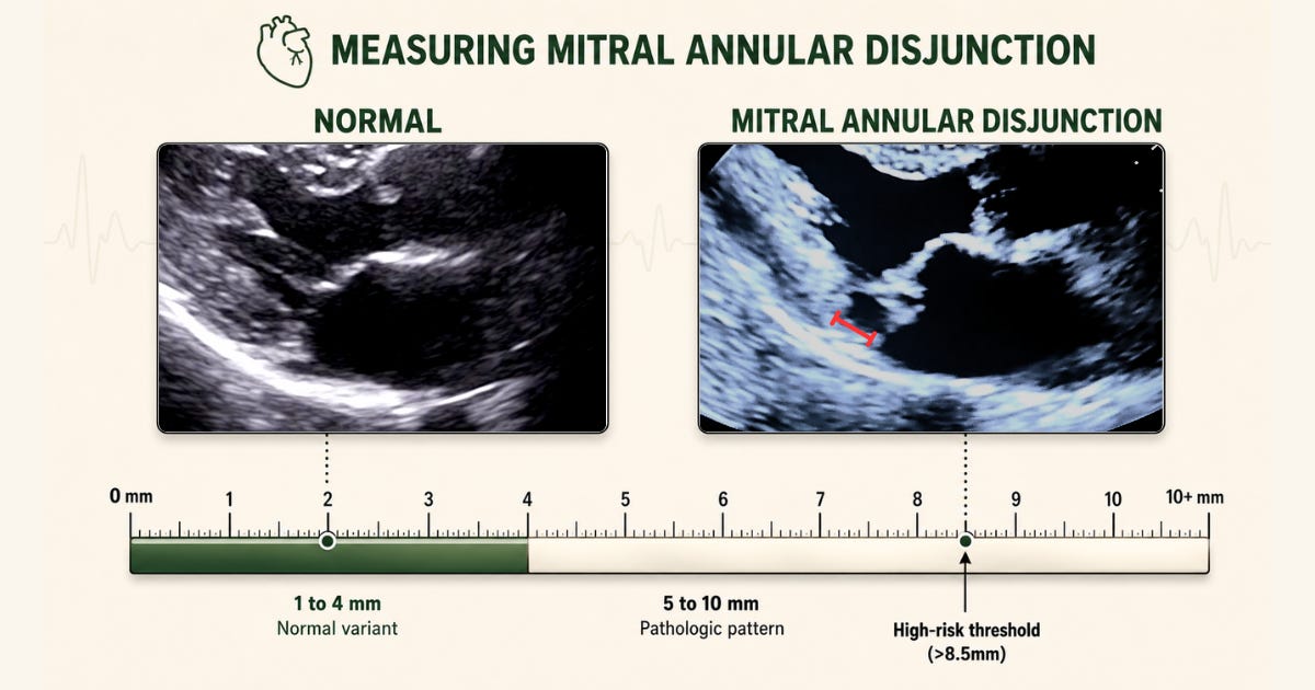
Take a look at the example below. Can you tell which phenotype is being shown before you keep reading?
How often MAD shows up depends heavily on what you are looking with:
General population: pooled prevalence of 8.7%
MVP or Barlow’s disease: pooled prevalence of 30.1%
Structurally normal hearts on CT: disjunction seen in 96%, median height just 3 mm
That last number is not a sign that everyone has pathologic MAD. Small, localized disjunction is a common anatomic variant. What actually tracks with arrhythmia is larger disjunction that wraps around the P2 scallop and coexists with myxomatous MVP.
Most patients with MAD are asymptomatic and found incidentally during a routine MVP workup. A minority present with palpitations, presyncope, or ventricular arrhythmia. In severe cases, MAD has been identified at autopsy in young, otherwise healthy patients after sudden cardiac arrest.
Echocardiography is the first-line imaging tool for any patient with known or suspected MVP, which makes it your first opportunity to catch MAD. It is less sensitive than cardiac MRI, but it is fast, available, and can be paired with tissue Doppler to pick up the arrhythmic risk markers described later in this article.
