

The Missing Tricuspid Valve
Tricuspid Atresia

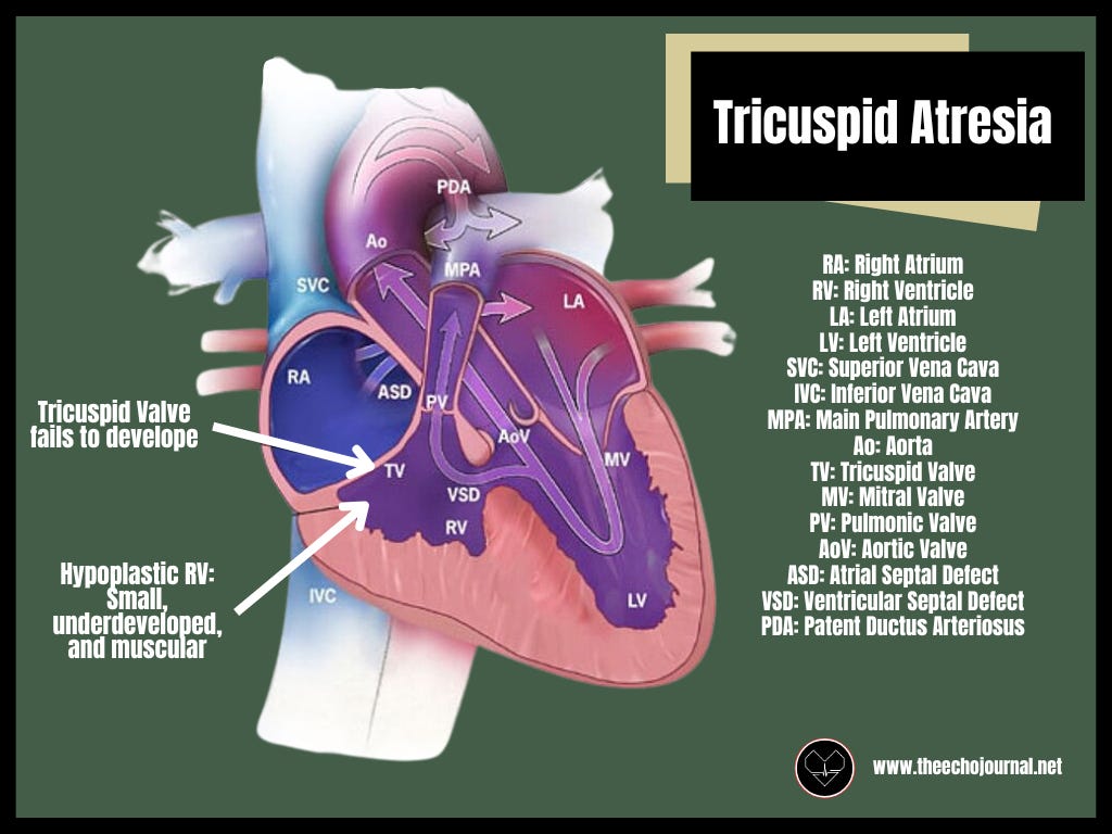
Tricuspid atresia is a rare but critical congenital heart defect where the tricuspid valve fails to develop, blocking direct blood flow from the right atrium to the right ventricle. This results in a hypoplastic right ventricle and necessitates alternative pathways for blood to reach the lungs. Affecting approximately 1 in 10,000 live births, tricuspid atresia accounts for about 1-2% of all congenital heart defects. Without surgical intervention, it carries a high mortality rate. However, with a staged surgical approach, many patients now survive well into adulthood with functional circulation.
Tricuspid atresia is classified into three types based on the relationship between the great arteries:
Type I (Normal Great Artery Relationship) 70% to 80%: The aorta arises from the left ventricle, and the pulmonary artery arises from the right ventricle. Pulmonary blood flow depends on a patent ductus arteriosus (PDA) or, if present, a ventricular septal defect (VSD).
Subgroup a: Intact ventricular septum with pulmonary atresia
Subgroup b: Small VSD with pulmonic stenosis (PS) or hypoplasia
Subgroup c: Large VSD without PS
Type II (Transposed Great Arteries) 12% to 25% : The aorta arises from the right ventricle, and the pulmonary artery arises from the left ventricle, similar to transposition of the great arteries (TGA). Pulmonary circulation depends on a VSD or PDA, often both.
Subgroup a: VSD with pulmonary atresia
subgroup b: VSD with PS or hypoplasia
Subgroup c: VSD without PS
Type III (Other or Complex Malformations) 3% to 6%: Encompasses more complex anomalies, such as truncus arteriosus or double outlet right ventricle, where the great artery arrangement deviates significantly from standard anatomy, further complicating surgical management.
(Minocha, 2024)
From an echocardiographic standpoint, tricuspid atresia presents a unique set of challenges, requiring a thorough assessment of compensatory shunts, ventricular function, and post-surgical anatomy. Understanding how to approach these studies effectively is necessary for both accurate diagnosis and long-term follow-up.