

The Aortic Valve, Deconstructed
Anatomy, the Origins of Aortic Regurgitation, and Why the Jet Direction Matters

You can grade aortic regurgitation all day long. But if you cannot say why the valve is leaking, the surgeon cannot tell whether it can be repaired or has to be replaced.
That answer lives in anatomy, not in a pressure half-time measurement.
In this lesson, you will walk the aortic valve from annulus to sinotubular junction, map every mechanism of AR onto the classification surgeons actually use, and learn why an eccentric jet hugging the anterior mitral leaflet is telling you something a central jet never will.
Trace-to-mild aortic regurgitation can be found in up to 15% of routine echocardiograms, the sonographer’s role transitions from simple detection to strategic quantification.
Accurate identification of the 0.5% who progress to moderate-to-severe AR is a clinical imperative, given that untreated severe AR more than doubles a patient’s mortality risk.
Pass boards on your first attempt. The Elite Study Plan gives you 1,200+ practice questions sorted by difficulty, 250+ flashcards for active recall, 10 study guides, and an explanation behind every answer — so you learn the concept, not just the correct letter.
Introduction
The aortic valve is made of three semilunar cusps attached to the aortic wall, and those attachments form part of the sinuses of Valsalva. The highest points of attachment at the commissures define the sinotubular junction. The lowest point of attachment where the aortic leaflets dip into the left ventricular outflow tract is called the nadir of the cusps, defining the annular plane.
The coaptation zone of the leaflets, called the lunulae, is fairly uniform in thickness. The exception is a slightly more fibrous area at the anatomic midpoint of each cusp known as the nodules of Arantius. On echocardiography, this portion of the leaflet can appear more echogenic even though it is normal.
Here is the concept that makes everything downstream easier: the aortic valve is a functional unit, not three isolated flaps. Annulus, cusps, sinuses, and sinotubular junction work together. Dilate any one of them and coaptation fails, even when the cusps themselves are perfectly normal.
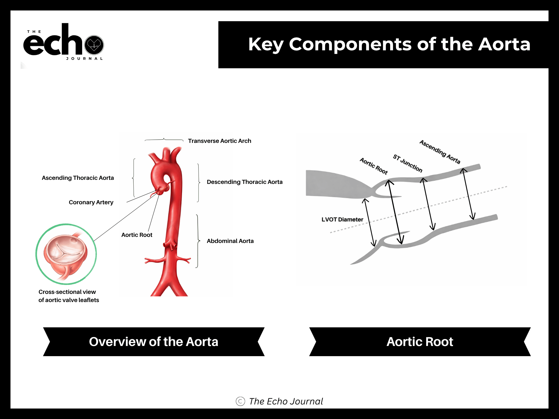
Where Aortic Regurgitation Comes From
Given that anatomy, AR results from disease of the aortic leaflets, the aortic root, or both, in each case producing valve malcoaptation. The 2017 ASE recommendations for evaluating native valvular regurgitation organize the causes this way.
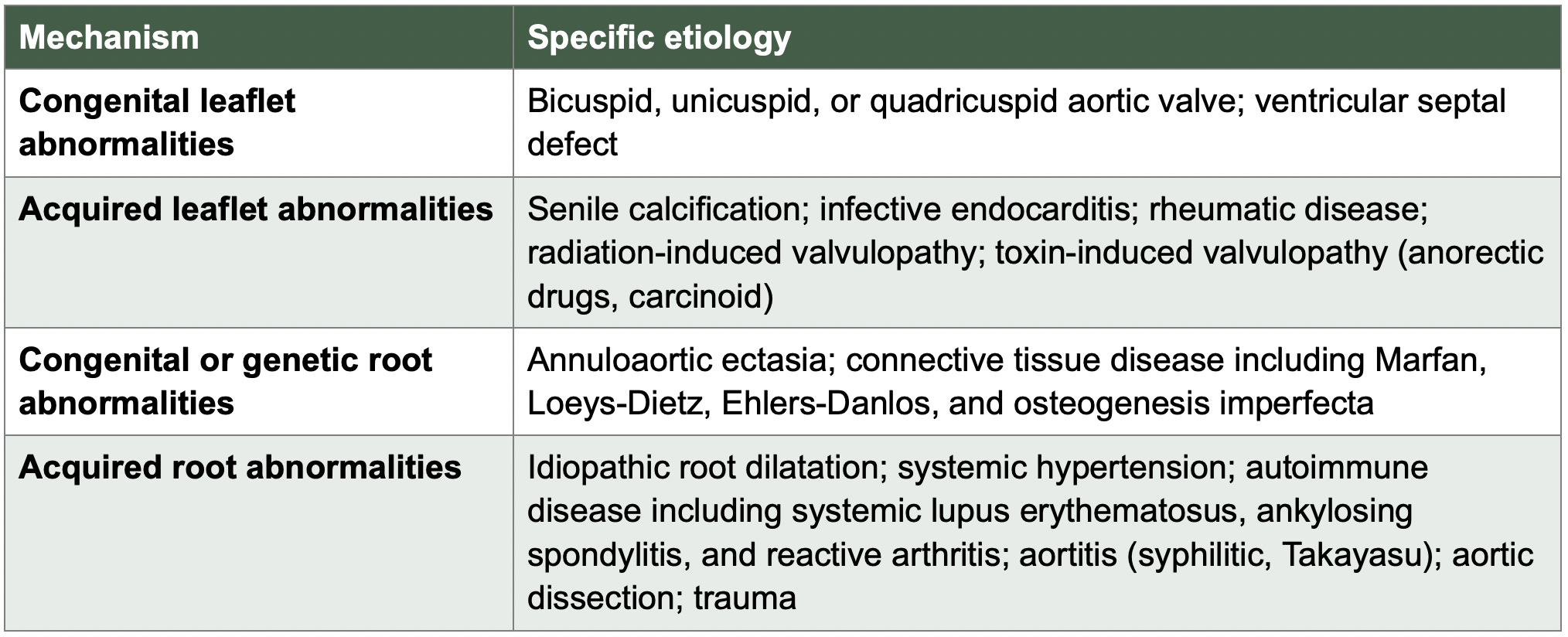
Bicuspid valves deserve a separate mention because you will see them constantly. All combinations of conjoined cusps can be identified on TTE, and visualizing the raphe is the key to classifying the type. Because stress is concentrated on the typically larger conjoined cusp, these valves can become stenotic, regurgitant, or both. AR may also appear secondary to the associated aortic dilatation rather than from the cusps at all. ASE reports TTE sensitivity up to 92 percent and specificity up to 96 percent for detecting bicuspid anatomy.