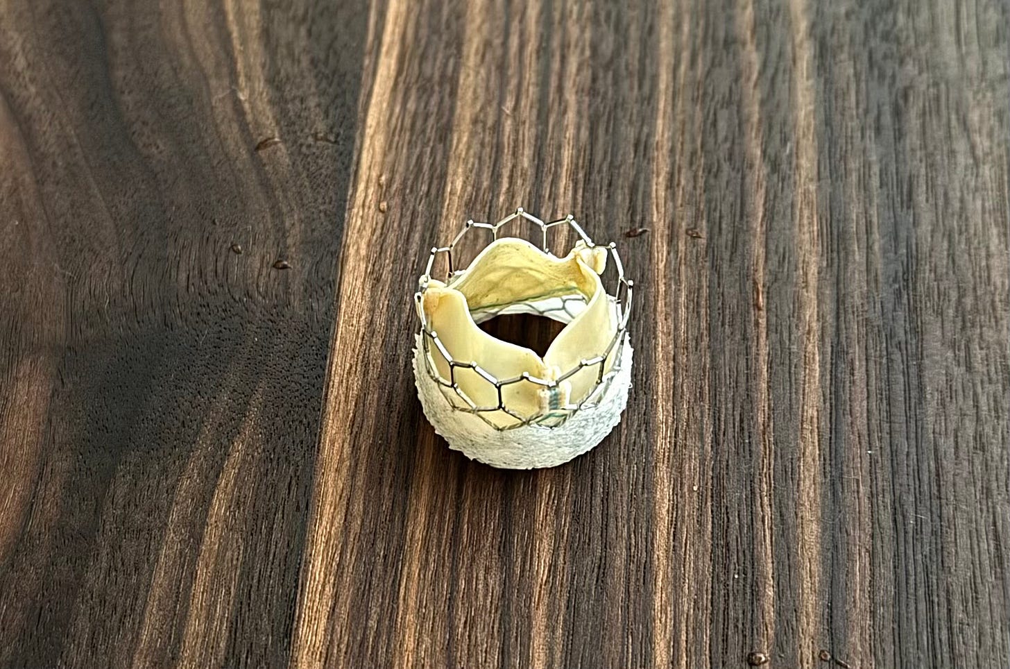
Transcatheter Aortic Valve Replacement (TAVR) has revolutionized the treatment of aortic valve disease, offering a lifeline to patients who might not survive traditional open-heart surgery. The role of a cardiac sonographer in the TAVR process is both critical and deeply rewarding, spanning preoperative evaluation, intraoperative guidance, and postoperative care. Using echocardiography, vital insights into the patient's heart can be provided, ensuring that every step of the procedure is as safe and effective as possible.
For more information about AS, please read:
History
In 1989, Doctor Henning Andersen speculated that expandable valves could be placed in a similar fashion to coronary stents. A few years later, he created a prototype for his idea that included a handmade metal stent, a porcine aortic valve, and a deflated balloon. He was unable to find a company that would bring his idea to fruition. French Cardiologist, Alain Cribier, pioneered the further development of TAVRs through the help of a company called, Percutaneous Valve Technologies. Animal experimentation took place for a couple years before the first successful human implantation in 2002 on a 57-year-old patient. Remarkable hemodynamic improvement proved the feasibility of TAVR and prompted rapid adoption of the technology in the coming years.


